Hypertension:
Persistent systolic pressure, more than 140 and diastolic more than 90 mm Hg. is regarded as hypertension.
The range of pressure increases with age.
Hypertension is the single most important risk factor in both coronary heart disease and cerebrovascular accidents.
It may also lead directly to congestive heart failure (hypertensive heart disease), renal failure and aortic dissection.
About 90% of hypertension is primary and idiopathic (essential).
The remaining is secondary and mostly related to renal disease or (less often) to renal artery stenosis (renovascular), endocrine abnormalities, vascular malformations, or neurogenic disorders.
Causes of hypertension:
I. Primary, essential, or idiopathic hypertension:
II. Secondary hypertension is due to the following causes:
1. Renal :
Acute glomerulonephritis ;
Chronic renal disease ;
Polycystic disease ;
Renal artery stenosis ;
Renal vasculitis ;
Renin-producing tumors ;
2. Endocrine:
Adrenocortical hyperfunction : Cushing syndrome, primary aldosteronism, congenital adrenal hyperplasia ;
Estrogen hormones : glucocorticoides, estrogen - including pregnancy and oral contraceptive, sympathomimetics, tyramine-containing foods, monoamine oxidase inhibitors ;
Pheochromocytoma ;
Achromegaly ;
Myxedema ;
Thyrotoxicosis ;
3. Cardiovascular :
Coarctation of aorta ;
Polyarteritis nodosa ;
Aortic insufficiency ;
Increased intravascular volume ;
Rigidity of the aorta ;
4. Neurologic:
Psychogenic ;
Increased intracranial pressure ;
Sleep apnea ;
Acute stress, including surgery ;
Regulation of Normal Blood Pressure :
Blood pressure is a complex trait that is determined by the interaction of multiple genetic and
environmental factors that regulate the relationship between cardiac output and total peripheral resistance.
1. Vasoconstriction increases vascular resistance.
Vasoconstrictors include angiotensin II, catecholamines, thromboxane, leukotrienes, and endothelin.
2. Vasodilators include kinins, prostaglandins, nitric oxide, and adenosine.
3. Regional autoregulation is also important, wherein increased blood flow leads to vasoconstriction and vice versa.
4. Cardiac output is regulated by blood volume (affected by sodium load, mineralocorticoids, and natriuretic factors), heart rate, stroke volume and contractility.
Mechanism of Essential Hypertension :
Although unknown, the cause of essential hypertension at the most elemental level must be related to a primary increase in cardiac output (e.g., reduced renal sodium excretion) or to an increase in peripheral resistance (e.g., owing to increased release of vasoconstrictor agents, to increased sensitivity of vascular smooth muscle cells, or to behavioural or neurogenic factors), or both (i.e., an increase in cardiac output and an increase in peripheral resistance).
In most patients, multiple defects probably contribute to the disease.
Abnormalities in the renal mechanisms that regulate blood pressure also may contribute to essential hypertension, including:
1. The renin-angiotensin system.
2. Sodium homeostasis.
3. Production of vasodepressor substances : It is implicated in the pathogenesis of hypertension in unilateral renal artery stenosis and renal disease.
For example, studies have suggested a propensity toward hypertension in individuals with specific molecular variants of the gene-encoding angiotensinogen, the physiologic substrate for renin.
Wednesday, August 29, 2012


congenital anomalies & arteriosclerosis
Congenital anomalies include the following conditions:
1. Anomalous (e.g. aberrant, reduplicated) vessels are principally of interest to surgeons.
2. Berry aneurysms are outpouchings of vessels as a result of congenital focal weakness.
They occur exclusively in cerebral vessels and occasionally rupture catastrophically.
3. Arteriovenous fistula is an abnormal communication between artery and vein.
It may be congenital or secondary to trauma, inflammation, or healed ruptured aneurysm.
1. Anomalous (e.g. aberrant, reduplicated) vessels are principally of interest to surgeons.
2. Berry aneurysms are outpouchings of vessels as a result of congenital focal weakness.
They occur exclusively in cerebral vessels and occasionally rupture catastrophically.
3. Arteriovenous fistula is an abnormal communication between artery and vein.
It may be congenital or secondary to trauma, inflammation, or healed ruptured aneurysm.
anatomy & physiology of the vascular system
Anatomy and Physiology of the Vascular System:
Arterial walls are thicker than veins.
Thickness of the arterial walls gradually diminishes as it becomes smaller.
Walls of the blood vessel consist of three basic structures, intima, media, and adventitia, which vary with the types of the vessels.
Blood vascular tree is a circuit that conducts blood from the heart through large- diameter, low-resistance conducting vessels to small arteries and arterioles, which lower blood pressure and protect the capillaries.
Capillaries are thin-walled and allow the exchange of nutrients and waste products between tissue and blood, a process that requires a very large area.
The circuit back to the heart is completed by the veins, which are disrensible and provide a volume biffer that acts as a capacitance for the vascular circuit.
Arteries are of three types:
1. Larger or elastic arteries (aorta and its main branches) :
Intima is composed of lining endothelial cells, separated from the media by internal elastic lamina.
Media is composed of smooth muscle cells.
Outer limit of the media is separated is separated from adventitia by external elastic lamina. Small arterioles (vasa vasorum) pass through the adventitia into the outer one-half to two-thirds of the media to perfuse the vessel wall.
Atherosclerosis is the disease largely of elastic and muscular arteries which is associated with functional and structural changes in the muscular arteries and arterioles.
2. Muscular arteries, are branches of elastic arteries (e.g. coronary or renal arteries) regulate the blood pressure by vasodilatation or vasoconstriction under the control of the autonomic nervous system.
3. Arterioles regulate the blood flow into capillary beds by the smooth muscle of media, thus controls the systemic arterial blood pressure.
Capillaries:Capillaries are small vessels having a diameter of a red blood cell, lined by one-cell thick endothelium, a thin basement membrane, and slow blood flow.
These are ideally suited to the rapid exchange of diffusible materials between blood and extravascular tissue.
Veins:
Veins are thin-walled vessels with poorly defined internal elastic lamina and media. Hence, they often show abnormal, irregular dilation, and easy penetration by cancer cells and inflammation.
Lymphatics:
Lymphatics are identified in tissue sections as collapsed, endothelium-lined channels devoid of blood cells.
Arterial walls are thicker than veins.
Thickness of the arterial walls gradually diminishes as it becomes smaller.
Walls of the blood vessel consist of three basic structures, intima, media, and adventitia, which vary with the types of the vessels.
Blood vascular tree is a circuit that conducts blood from the heart through large- diameter, low-resistance conducting vessels to small arteries and arterioles, which lower blood pressure and protect the capillaries.
Capillaries are thin-walled and allow the exchange of nutrients and waste products between tissue and blood, a process that requires a very large area.
The circuit back to the heart is completed by the veins, which are disrensible and provide a volume biffer that acts as a capacitance for the vascular circuit.
Arteries are of three types:
1. Larger or elastic arteries (aorta and its main branches) :
Intima is composed of lining endothelial cells, separated from the media by internal elastic lamina.
Media is composed of smooth muscle cells.
Outer limit of the media is separated is separated from adventitia by external elastic lamina. Small arterioles (vasa vasorum) pass through the adventitia into the outer one-half to two-thirds of the media to perfuse the vessel wall.
Atherosclerosis is the disease largely of elastic and muscular arteries which is associated with functional and structural changes in the muscular arteries and arterioles.
2. Muscular arteries, are branches of elastic arteries (e.g. coronary or renal arteries) regulate the blood pressure by vasodilatation or vasoconstriction under the control of the autonomic nervous system.
3. Arterioles regulate the blood flow into capillary beds by the smooth muscle of media, thus controls the systemic arterial blood pressure.
Capillaries:Capillaries are small vessels having a diameter of a red blood cell, lined by one-cell thick endothelium, a thin basement membrane, and slow blood flow.
These are ideally suited to the rapid exchange of diffusible materials between blood and extravascular tissue.
Veins:
Veins are thin-walled vessels with poorly defined internal elastic lamina and media. Hence, they often show abnormal, irregular dilation, and easy penetration by cancer cells and inflammation.
Lymphatics:
Lymphatics are identified in tissue sections as collapsed, endothelium-lined channels devoid of blood cells.
Monday, August 27, 2012
SCHISTOMIASIS (S.JAPONICUM, S. HAEMATOBIUMHISTOSOMA MANSONI)
Schistomiasis
 |
| s. japonicum male |
Schistosomes are among the most important blood parasites of man. Human schistosomiasis (bilharzia or bilharziasis) is due to three species of , ScSchistosoma: Schistosoma haematobiumhistosoma mansoni and Schistosoma japonicum. S. haematobium is responsible for urinary schistosomiasis in Africa , and the Middle East ; S. mansoni is responsible for intestinal schistosomiasis in Africa , Central and South America and the Caribbean and S. japonicum causes intestinal schistosomiasis in the Far East , particularly China Japan Philippines
Although the three major human schistosomes are primarily parasites of man, S. japonicum is present in many other mammals including cattle, goats, pigs, dogs and cats. These animals serve as reservoirs from which infection can be passed to man. The role played by wild animals in human infections of S. haematobium and S. mansoni has not been clarified.
The schistosomes are digenetic trematodes with relatively complex life cycles. The male that measures about 8 to 16 mm long has a groove, the gynecophoric canal, in which the longer and more slender female is held. The male and the female are in permanent copula and usually live for many years.
Life cycle
The adult worms live in the small mesenteric veins of the small intestine or pelvic veins where the females deposit their eggs. Each female S. japonicum lays about 1300 eggs daily. This is about four times the number of eggs shed by the female S. mansoni or S haematobium, which are around 350 eggs per day.
Each species of Schistosoma has characteristic eggs. The eggs of S. haematobium have a large terminal spine; those of S. mansoni have a lateral spine while those of S. japonicum have a rudimentary lateral spine that is often difficult to locate.
As the eggs are laid, they work their way out of the small blood venules aided by their own hydrolytic secretions. The eggs of S. japonicum and S. mansoni reach the lumen of the small intestine by traversing the intestinal musculature before being expelled in faeces. Those of S. haematobium move from the surrounding pelvic blood vessels into the bladder and pass out in urine.
Some of the eggs are carried to the liver and lungs where they cause serious inflammatory tissue reactions, which usually culminate in the formation of fibrous tissue around them.
 |
| S. JAPONICUM FEMALE |
The shelled eggs are fully developed embryos and hatch into free - swimming ciliated miracidia upon contact with water of low salt content. The miracidia survive for up to 24 hours. For continuation of life, the miracidium must penetrate a snail host during this period. On finding a suitable snail, the ciliated epithelium is shed, the larva becomes a tubular sporocyst that grows to about 1 mm in length and for the next two to three weeks produces daughter sporocysts. Daughter sporocysts emerge from the mother sporocyst, undergo growth and migrate from the snail’s viscera to the digestive gland.
The larvae are very specific in their choice of snails. S. japonicum miracidia can only penetrate the tissues and establish in freshwater snails of the genus Oncomelania, while S. mansoni will only invade snails of the genus Biomphalaria, and S. haematobium those of the genus Bulinus.
In the molluscan digestive gland, sporocysts divide repeatedly into daughter sporocysts, first and second-generation rediae. About six weeks since the miracidium entered the snail, fork – tailed cercariae emerge from the snail. The cercaria is a free-swimming organism that must enter the skin of a mammalian host within two to three days or it dies.
During the penetration of the skin of a mammalian host, the cercariae shed their tails and within a few hours, the young flukes, called schistosomulae, migrate through veins and lymph vessels to the lungs. From there they migrate to the liver, where they develop into young male and female worms within portal blood vessels.
After 4-6 weeks, mating takes place and worm pairs move to their final destinations, which, in urinary schistosomiasis, are the blood vessels of the bladder, and intestinal mesenteries in the case of S. japonicum and S. mansoni. Thereafter, the female worms begin shedding eggs. Some of the eggs make their way through the walls of the blood vessels and enter the bladder or the intestinal lumen. Those of S. haematobium pass out in urine, while those produced by the two intestinal schistosomes are voided in faeces.
Pathogenesis
The main pathological features associated with schistosomiasis are mechanical damage caused by the movement of the eggs through the venules to the intestine or urinary bladder, the formation of fibrotic tissue around the eggs and inflammation. Large numbers of eggs cause extensive rupture of the membranes of the bladder and intestine, resulting in haemorrhage and blood in urine and faeces. About 50 % of the eggs produced by the female worms are trapped in the tissues. Depending on the species of Schistosoma, the clinical manifestations involve inflammatory reactions due to eggs lodged in the liver, intestinal and urinary systems.
Urinary schistosomiasis is characterised by painful urination, and progressive damage to the bladder and ureters. This in turn leads to narrowing of the urinary tract and obstruction of urine flow. In serious cases, there may be total kidney failure.Bladder cancer is common in advanced cases.
 |
| S. JAPONICUM ADULT MALE+ FEMALE |
The pathology of S. mansoni and S. japonicum is largely similar. However, tissue damage in the Oriental parasite is more severe mainly because of the large numbers of eggs it releases. The fact that S. japonicum is also a parasite of many kinds of mammals seems to suggest that it could have started as an animal parasite that later adapted to man. That being the case, it explains why it is normally considered more virulent than the other two species of human schistosomes.
The damage to the liver and intestinal walls is common and is caused by the fibrotic lesions that develop around eggs trapped in these organs. Long-term chronic infections eventually lead to hypertension of the abdominal blood vessels and progressive enlargement of the liver and spleen.
Long-standing portal hypertension causes secondary enlargement of other vessels such as the superficial veins of the abdominal wall, particularly varicose oesophageal veins from which bleeding can be fatal.
Symptoms
During the early stages of infection, patients with S. japonicum or S. mansoni experience tiredness, recurrent afternoon fever, night sweats, diarrhoea, loss of appetite and loss of weight. The symptoms due to S. haematobium at this stage are not very different from those of the other two species. The patient complains of fever, moderate hepatic and epigastric pain. More often, the obvious symptoms of infection are the presence of blood in the urine and a sharp pain during micturation.
The usual means of human exposure to infection consists of wading, swimming, and bathing or washing clothes in shallow freshwater along the banks of rivers, lakes and ponds infested with infected snails. Urine and excreta deposited by infected persons that find their way to the water are the major source of infection for the snails. Reservoir animals in S. japonicumcontribute to the pollution of water with their infected faeces and increase the chances of human infection.
Schistosomiasis may well be described as an occupational hazard in as far as it affects mostly paddy rice farmers and fishermen. Other groups of people that are usually more exposed to infection than the rest of the population because of their nature of work are women and children. In rural areas, women spend a lot of time in contact with water as they wash family clothing and fetch water for domestic use. Likewise, young children spend a lot of their time swimming and playing in water, thereby exposing themselves to infection.
As the human population increases, there is a corresponding increase in the demand for food. To meet this demand, farmers turn to damming rivers to create large water reservoirs for irrigation. More fishponds are dug and swamps are exploited for rice and other agricultural production. These activities create favourable conditions for the snails to multiply and increase the transmission and prevalence of schistosomiasis.
 |
| S. JAPONICUM OVA |
The construction of the Aswan Egypt Ghana Zambia Nigeria Mali
 | |
|
A few wild animals have been found naturally infected with S. haematobium but their role in human infection is unclear. However, rodents, baboons and velvet monkeys are known to harbour of S. East Africa . Whether these animals are natural reservoirs of human infection remains to be resolved.
Diagnosis
Examination of the faeces reveals characteristic eggs of S. mansoni or S. japonicum. For S. haematobium, the eggs are detected in urine.
Control Methods
 A successful control method will have to take into account provision of piped water, wearing of protective gear by fishermen and farmers, use of toilets and avoidance of swimming in infected waters.
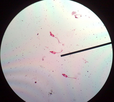 Snail intermediate hosts are controlled using molluscicides, applied along river and lakeshores and along irrigation canals. Although molluscicides are effective in limiting infection, most of them are unfortunately quite toxic and end up killing many other nontarget organisms including the fish.
| S. JAPONICUM |


 |
| S. MANSONI OVA |
Drugs used for treatment of schistosomiasis include metrifonate, oxamniquine and praziquantel. Metrifonate is cheap but requires three doses and is only effective against S. haematobium. Oxamniquine is effective as a single dose against S. mansoni. Praziquantel is a safe, effective drug against all species of Schistosoma and is given as a single dose. Treatment with praziquantel is followed by a dramatic decrease in parasite numbers and egg production. A reduction in parasite numbers means there are fewer eggs, which means fewer snail intermediate hosts will be infected. Eventually, there is a decrease in the transmission of infection. Praziquantel is also effective against trematodes and cestodes.
Sunday, August 26, 2012


Subscribe to:
Posts (Atom)