Balantidium coli
The ciliate protozoa form a large group of organisms that is characterised by numerous cilia. A large number of ciliates are free-living and an equally large number consists of parasites of the digestive systems of termites, cockroaches and herbivores.
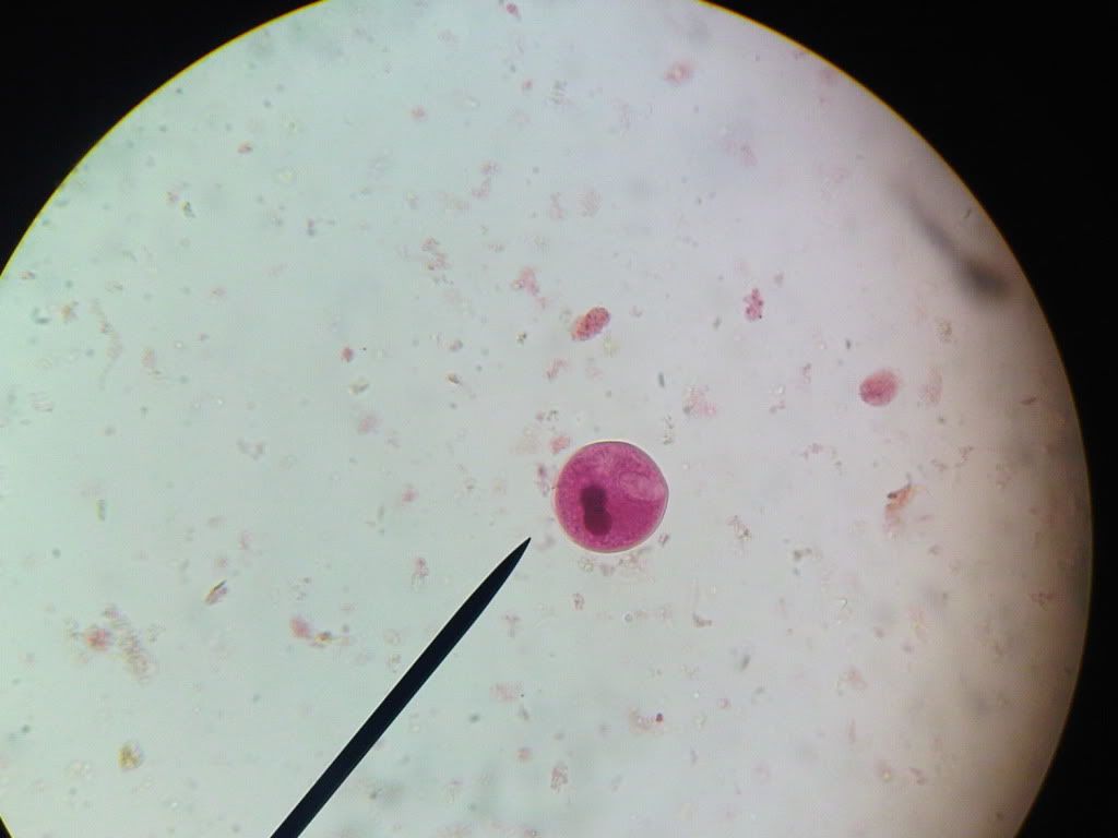
A typical ciliate has a conical mouth, the cytostome, near the anterior end and an anal opening, the cytopyge, at the posterior end. Ciliates have two types of nuclei: a large kidney-shaped macronucleus and a small spherical micronucleus, both of which are visible in the cyst and in the trophozoite.

Balantidium coli is the only ciliate that is parasitic to humans. Balantidium coli has a global distribution in pigs, dogs and several species of monkeys. In man, it occurs mainly in warm climates. The parasite occurs in the large intestine of man, pigs and monkeys.
Morphology
The parasite exists as a cyst and as a trophozoite, the trophozoite being the active form. It is ovoid and covered by cilia that are perpetually in motion, which are used for movement. The parasite varies in size, ranging from 50 to 100 µm in length by 40 to 70 µm in width. The anterior end is conical and the posterior end is rounded. To one side of the anterior end, there is funnel-like peristome, which leads to the cytostome or mouth.


Balantidium coli trophozoite (left) and cyst (right)
Life cycle
Balantidium coli lives in the large intestine where it feeds on bacteria, cells and food particles present in the intestinal lumen. Asexual reproduction is by binary fission in which the micronucleus first divides followed by the division of the macronucleus, resulting in two daughter organisms. B. coli can also reproduce sexually by a process called conjugation, which involves the exchange of nuclear material.

Life cycle of B. coli. A, ingestion of cyst; B, trophozoite formed in the colon divides by binary fission;
C, trophozoite in the lumen of the large intestine or in faeces freshly voided.
Pathogenesis
The effect of the infection in the pig is minimal, as the parasite does not seem to penetrate the mucosal surface. In humans and monkeys, the parasite can enter the surface of the large intestine causing extensive tissue erosion. Furthermore, the parasite may penetrate through the muscularis mucosae into the submucosa and spread out radially causing rapid destruction of the tissues. Most of the lesions caused by Balantidium coli are found in the caecal and sigmoid rectal regions.
The symptoms vary from severe diarrhoea and dysentery to mild insignificant, almost asymptomatic condition.
Epidemiology

Infection is acquired by the ingestion of cysts of Balantidium in food or water contaminated with faecal matter. Since balantidiasis is common in dogs and primates, it may be expected that these animals are probably the main source of infection to man. However, human infections are usually associated with people who come in contact with pigs, chiefly pig keepers or butchers.
The infection rate is very low in humans and this is because humans are generally refractory to exposure to Balantidium. However, once the infection has established itself in man, there is usually a rapid transmission from one person to another, particularly where there is no proper sanitation.
Diagnosis and treatment
Slide smears of infected faeces should show characteristic cysts or trophozoites of Balantidium coli. The infection responds to treatment with tetracycline, and metronidazole.
. Apart from these, they also provide information related to community mental health services designed specifically for children, refugees, HIV infected patients, the elderly, etc. Here are a list of various community health services available for your use. can diabetics have pizza hut
ReplyDeleteThank you for taking the time to publish this information very useful! Medical Ancillary Services
ReplyDelete